Your on-line source for reliable and unbiased information about the evaluation & treatment of heart disease.
First | 1 | 2 | 3 | 4 | 5 | 6 | Last
The Cardiac Cath learning module is made up of 6 parts or sections. You may navigate through the pages by clicking on the green arrows or numbers (above), the specific questions (below) or the gray menu items on the left.
What is Cardiac Catheterization?
What Preparations are Needed?
How is Cardiac Cath performed?
What do I need to know about the equipment?
What is experienced in the Cath lab?
How long does it take?
What preparations are needed?
What happens after arrival in the cath lab area?
How safe is the procedure?
What is the reliability of the test?
How quickly will I get the results?
Show me a panoramic view of the Cardiac cath lab?
What is Cardiac Catheterization?
Cardiac Catheterization (Cath) is a specialized study of the heart during which a catheter, or thin hollow flexible tube, is inserted into the artery of the groin or arm. Under x-ray visualization, the tip of the catheter is guided to the heart. Pressures are measured and an x-ray Angiogram (Angio) movie of the heart and blood vessels are obtained while injecting an iodinated colorless "dye" or contrast material through the catheter. Coronary angios are obtained by injecting the contrast material into the opening or mouth of a coronary artery. The iodinated solution blocks the passage of x-rays. X-ray movie pictures taken during the injection of the contrast material allow the coronary arteries to be visualized. In other words, coronary arteries are not visible on x-ray film. However, they can be made temporarily visible by filling the coronary artery with a contrast solution that blocks x-ray.
The coronary arteries are vital because they supply oxygen and nutrients to the heart muscle. Without blood flow, the muscle would sustain permanent damage in the form of a heart attack or myocardial (pronounced my-ow-card-yull) infarction (pronounced in-fark-shun).
Cardiac Catheterization (Cath) is also known as Heart Cath, Angiogram (Angio) (pronounced an-gee-o-gram) or Arteriogram (pronounced ar-tee-rio-gram). The latter two terms describe the use of contrast material to take x-ray pictures of the heart.
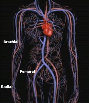
If catheters are introduced through the femoral (pronounced fem-rull) or groin artery, the procedure is known as "left heart" catheterization, because the catheter goes from the femoral artery to the aorta, coronary arteries, and the Left Ventricle (LV). This accounts for the majority of procedures. Left heart cath can also be performed by using the artery in the arm.
If a catheter is also placed in the right femoral vein to measure pressures within the right side of the heart, the procedure is called "right heart" catheterization. This is used in patients with congenital heart disease, diseases of the heart valve, or certain conditions involving the pericardium (pronounced perry-card-e-yum), or sac, of the heart. This may also be used in certain diseases of the heart muscle, heart failure, shock, or when measurements of heart output or lung pressures are needed. Right and left heart catheterization is a combination of both.
![]()

There are three major arteries that run on the surface of the LV. This is the most important pumping chamber of the heart and supplies oxygenated blood to the body. The aorta arises from the LV and gives out a series of branches as it makes its way from the heart to the lower portion of the abdomen. The coronary arteries are the very first branches that arise from the aorta. There are two major coronary branches that come off the aorta. The one that arises from the left is known as the Left Main Coronary Artery. This immediately divides into the Left Anterior Descending (LAD) and the Circumflex (Circ).
The LAD supplies blood to the front portion (anterior wall) of the LV and the septum (partition wall that separates the LV from the Right Ventricle (RV)).
The Circ wraps around the heart and supplies blood to the back or posterior wall of the LV.
The Right Coronary Artery (RCA) arises from the right side of the aorta and supplies blood to the bottom or inferior wall of the LV. It also supplies branches to the RV.
After giving rise to the coronary arteries, the aorta continues down towards the belly and gives off branches that go past the groin and into the legs. The cardiologist can insert a catheter or thin plastic tube into the groin artery (where it comes close to the skin) and "thread it" UP the aorta and to the mouth or openings of the coronary arteries. X-ray is used to guide the passage of the catheter. As noted earlier, an iodinated contrast solution is then injected into the coronary arteries to make it visible on the x-ray. A video image and movie film are simultaneously recorded during injection into the coronary artery. Below is a panoramic view of the cath lab.
Panoramic View of the Cardiac Cath Lab
![]()
©1999-2017, 20XXA.S.M. Systems, Inc. All Rights Reserved, including design and all graphic contents & animations