Your on-line source for reliable and unbiased information about the evaluation & treatment of heart disease.
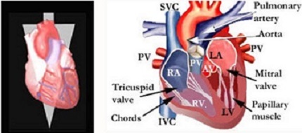 The various portions of the heart is shown in the adjoining images.
The image on the right shows a longitudinal (cut from top to bottom) section of the heart while the image on the left shows the plane through whick the section was taken. Oxygenated blood from the lungs is returned to the heart through four pulmonary veins (PV) that empty into the left atrium (LA). When the left atrium is filled completely, the pressure that it creates opens the mitral valve (MV) very much like opening a hatch door by applying pressure against it. The edges of the mitral valve that is made up of two leaflets are kept from flopping back into the left atrium by cords that are known as chordae tendineae (pronounced cord-ee ten-din-ee). This is very much like ropes that holds the sail of a boat in place as wind gushes against it. The other end of the cords are attached to papillary muscles that helps take up the slack as ventricle starts to empty (known as contraction) and decreases in size. The increased pressure within the ventricle as it contracts pops open the aortic valve and blood gushes into the aorta. The aorta serves as a highway or conduit that divides into branches which in turn carries blood to various parts of the body.
The body tissues extracts oxygen and nutrients from the blood that is then returned to the right atrium (RA), travels through the tricuspid valve and enters the right ventricle (RV). The process is very similar to that described for the left atrium and left ventricle. Unlike the mitral valve that has two leaflets, the tricuspid valve has three leaflets (thus the name). When the right ventricle (RV) is filled, it begins to contract and empties the blood across the pulmonary valve and into the pulmonary artery (PA). The PA carries blood to the lungs where it picks up oxygen. The oxygenated blood then travels via the pulmonary veins and into the left atrium. This round trip comprises the circulation of blood.
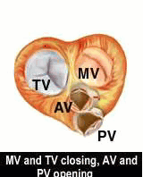
The pictures below represent a heart that is cut along the horizontal axis. The picture on the left shows the plane along which the heart is cut. That is, the top of the heart, including the right and left atria (atria is plural for atrium), the pulmonary artery and aorta are removed on the picture on the right (below). It shows the heart as you would look down at it from the front. The tricuspid and mitral valves are represented right and left, respectively (you can see the right and left ventricles through the two valves). The aortic and pulmonic valves are shown up and down, respectively, in the bottom half of the picture. The heart size increases and decreases during the filling (DIASTOLE, pronounced die-as-tull-ee) and contraction or emptying (SYSTOLE, pronounced sis-tull-ee) of the heart chambers.
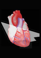
 The animation on th right shows an animated cross section of th heart, together with valve structures that open and shut to let blood pass through the atria, ventricles and the great vessels. The image on the left shows the plane through which the cross-section of the heart was taken.
The video clip below shows another cross section of the heart.
.
The mitral and tricuspid valves open and the aortic and pulmonic valves are shut while the ventricles fill during diastole. In contrast, the mitral and tricuspid valves shut while the aortic and pulmonic valves open during ventricular systole. This sequence ensures that the ventricles are filled to capacity before the aortic and pulmonic valves are opened. At this time, the mitral and tricuspid valves are shut so that blood does not leak back into the the two atria. Yes, the heart is an ingenious device that could have inspired design of the modern day mechanical pump and integrated valves.
Did you stop and wonder why each side of the heart has two pumping chambers (atrium and ventricle)? Why not just have a ventricle to receive blood and then pump it straight out? The reason is that the atrium serves as a "booster pump" that increases the filling of the ventricle. Filling a normal ventricle to capacity translates to more vigorous contraction or emptying. You can compare this to a strong spring, and imagine that the heart muscle is made up of tiny little "springs" known as ACTIN and MYOSIN. Within reasonable limits, the more you stretch a spring, the more vigorously will be its contraction or recoil. In medical terms, this is known as "Frank-Starling's" law.
![]()
![]()
©1999-2017, 20XXA.S.M. Systems, Inc. All Rights Reserved, including design and all graphic contents & animations