Your on-line source for reliable and unbiased information about the evaluation & treatment of heart disease.
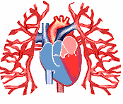
Let us now follow the circulation of blood through the heart. As noted earlier, oxygenated blood is pumped by the left ventricle to all parts of the body, other than the lungs. The body tissue removes much of the oxygen for its own need. The blood, which is now carrying less oxygen, returns to the heart. Blood from the head, neck and arms return to the right atrium (RA) via the SVC or SUPERIOR VENA CAVA. On the other hand, blood from the lower portion of the body returns to the RA via the IVC or INFERIOR VENA CAVA (pronounced vee-nah cave-ah).
The RA contracts when filling is completed. This builds up pressure within that chamber and pushes the tricuspid valve open. Blood now rushes from the RA to the right ventricle (RV). When the RV is filled, the walls begin to contract and raises pressure within the RV. The increased pressure shuts the tricuspid valve and pumps blood into the pulmonary (pronounced pull-mun-narey) artery through the pulmonic valve (PV, pronounced pull-mon-nick) which is pushed open by the increased pressure. The diagram below once again shows the four heart valves as viewed from the top, standing in front of the heart, i.e., we are looking down at the two ventricles with the right atrium and left atrium removed.
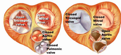
The pulmonic valve is made up of three cusps or flexible cup like structures. When the pressure in the right ventricle is low (as is the case during the filling phase of the chamber) the three cusps are full of blood and their sides touch each other to close the opening. This prevents blood from leaking into the pulmonary artery while the RV is filling.
When the RV contracts to empty, the pressure within the chamber rises above that of the pulmonary artery. This forces open the three cusps of PV and blood rushes through the pulmonary arteries and is sent to the lungs. Here the red blood cells pick up oxygen
The oxygenated blood from the lungs now returns to the left atrium (LA) via four tubes that are known as pulmonary veins. They empty into the back portion of the LA. When the LA contracts after it is completely filled. This opens the mitral valve and forces blood into the left ventricle (LV).
When the LV is completely filled, it starts to empty its contents by contacting the walls. This increases pressure within the chamber, shuts the mitral valve and opens the aortic valve (AV, pronounced a-ortic). The sequence is similar to that described for the RA, RV and pulmonic valve.
![]()
Blood now rushes through the aorta (pronounced a-or-tah). The aorta is the main "highway" blood vessel that supplies blood to the head, neck, arms, legs, kidneys, etc. Thus, blood is brought to each of these organs and limbs via branches that originate from the aorta. The cells within each part of the body pick up oxygen and nutrients from the blood. The oxygen-poor blood then returns to the RA, via the superior and inferior vena cava, and the beat goes on!!
The animation above demonstrates the flow of blood through the heart and lungs, as explained above. Notice that the mitral and the right side of the heart works in synchrony with the left, but that each atria contracts while the ventricle fills
Less confused? Good! Continue to hang in there as we further clarify these concepts
![]()
![]()
©1999-2017, 20XXA.S.M. Systems, Inc. All Rights Reserved, including design and all graphic contents & animations