Your on-line source for reliable and unbiased information about the evaluation & treatment of heart disease.
Inspection
Palpation or "hands-on" examination
Percussion or "tapping" examination
Auscultation or use of stethoscope
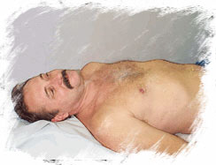
Inspection of the neck veins and its prominence could be indicative of heart failure and an excessive load on the right side of the heart. A bluish discoloration of the tongue and nail beds could point to a low oxygen level in the blood, while pallor or a pale appearance could indicate a low level of hemoglobin. Additionally, inspection of the chest may provide information about enlargement of the heart. Thus, a physician obtains an enormous amount of information even before touching the patient.
![]()
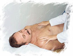
During palpation, the physician uses his or hands to examine the patient. During this phase, the physician can feel the heart beat and diagnose enlargement. Loud heart murmurs may also be felt without the use of a stethoscope. This is known as a "thrill." Palpation of the belly could help diagnose liver enlargement, find the tenderness of an active ulcer, or help uncover an aneurysm.
The patient's pulses are also felt to help determine if there is disease of the blood vessel accounting for calf pain when the patient walks. Pressing the legs and feet with the fingertip can diagnose the presence of edema or excess fluid.
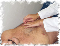
During percussion, the examiner places one hand on the patient and then taps a finger on that hand, with the index finger of the other hand. Since hollow and solid areas generate different vibrations, the physician or other examiner uses this technique to determine if various organs (heart, liver, etc.) are enlarged or not. Percussion is also used to diagnose fluid in the abdominal and chest cavities or make one suspect the presence of pneumonia.
![]()

Auscultation or listening with a stethoscope: During auscultation, the physician listens to the patient's heart beat, lungs and blood vessels of the neck and groin. Abnormal heart sounds, known as gallops, are a clue to heart disease. Also, the location, character and timing of a heart murmur (this is a prolonged sound that is created by turbulent blood flow across heart valves) are used to diagnose various valve diseases. However, it should be recognized that murmurs may also be heard in many normal individuals.
Certain characteristics of the murmur and other portions of the examination help the physician diagnose specific forms of heart diseases. Similarly, blockages in the arteries of the neck and those that supply the legs may also produce a turbulent flow. This can be heard with a stethoscope and is known as a "bruit" (pronounced broo-ee). Listening to the lungs, when integrated with the history and other portions of the physical examination, can diagnose such conditions as heart failure, accumulation of fluid, asthma, bronchitis, pneumonia, collapsed lungs, etc.
![]()
![]()
©1999-2017, 20XXA.S.M. Systems, Inc. All Rights Reserved, including design and all graphic contents & animations